
Vol. 11 No. 4 (2017): July
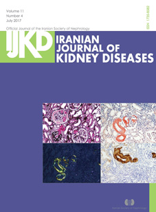
About the cover:A, Light microscopy showing nodular mesangial expansion and arteriolar thickening with silver-negative materials. B, Congo-red stain showing pink-reddish mesangial and vascular staining. C, A section showing typical apple-green birefringence under polarized light microscopy. D, Immunohistochemical staining for serum amyloid A is positive.
Published:
2017-08-10